badara :

Morphology:
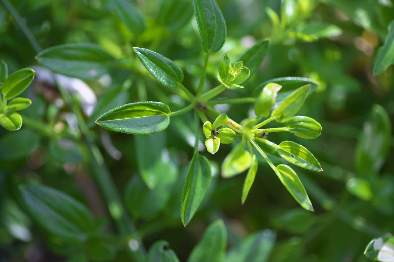
Spiny, deciduous shrub or a small tree, up to 10 m high;spines in groups of two, one straight, up to 2.5 cm long and one curved. Leaves alternate, petiolate, oval-lanceolate, 2–7 cm long, 2.5–3.0 cm wide;apex slightly obtuse;base oblique;margin closely serrulate, with three veins. Inflorescence an axillary cyme. Flowers perfect, seven to eight in each cluster;calyx with cupuliform tube and five segments;petals five, yellow;disk`lining the calyx tube;stamens five;ovary depressed into the disk.
Fruits are fleshy drupes, ovoid or oblong, 1.5–5.0 cm long, dark reddish brown when ripe. Irregular furrowed stones are found in tuberculate seed which contains 6 mm long brown kernels of elliptic shape.
Histology:
The longitudinal sections (L.S) of the fruit show thin green epicarp, wide soft mesocarp, and dark brown thick endocarp. In longitudinal view, the seed shows a thick conical part, thick wide shell which is hard. In the cotyledon is more or less cordate,white and soft with shallow notch at the upper end. In T.S, the fruit appears circular with soft pericarp, thick dark brown seed coat of sclereids, and vertically elongated white cotyledon. The epicarp layer is broken at certain places. In the mesocarp, some of the cells have dense tannin content and others have mucilage substance. The mucilaginous canals are wide, unbranched, and wavy. Some of the mesocarp cells contain dense accumulation of protein bodies. The sclerotesta contains palisade or macrosclereids. The seed consists of two elliptical, flat cotyledons which show dense accumulation of starch grains and small less prominent vascular strand. The powder microscopy of the fruit contains abundant dark mucilaginous substance. Fragments of epidermal cells of the pericarp and mesocarp cells are frequently seen. The seed coat epidermal cells are polyhedral with lignified cell walls. The circular brachy sclereids were often seen in the mesophyll tissue of the fruit. The ground parenchyma with various shapes and size is also noticed.- » Classification and names of badara
- » Synonyms and definitions of badara
- » Drug Properties of badara
- » Chemical Constituents of badara
- » Standardization of badara
- » Parts used and Dosage of badara
- » Morphology and Histology of badara
- » Distribution and Conservation of badara
- » Cultivation of badara
- » badara in the market
- » Medicinal Uses of badara
- » Researches and clinical trails of badara
- » badara in other sytems of medicine
- » Ayurvedic formulations with badara
- » Images of badara
Website Administrator
This article is incomplete. If you feel you can make a contribution, please let us know by sending email to mail@ayushvedah.com, we may consider your request. Thank you.